PlantScreen Imaging sensors
PlantScreen Imaging sensors
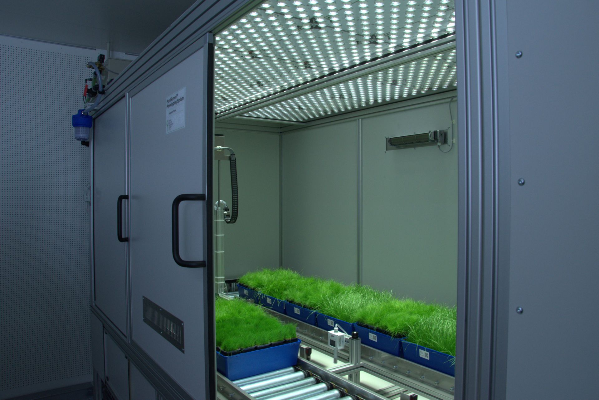
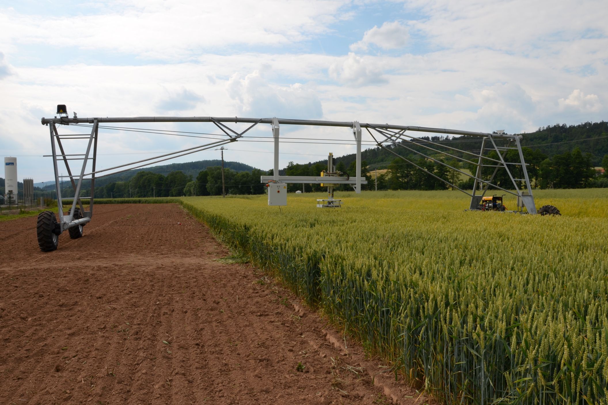
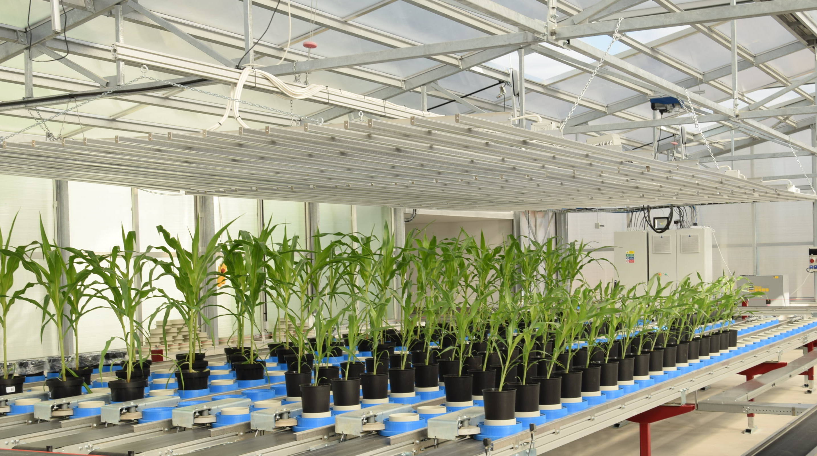
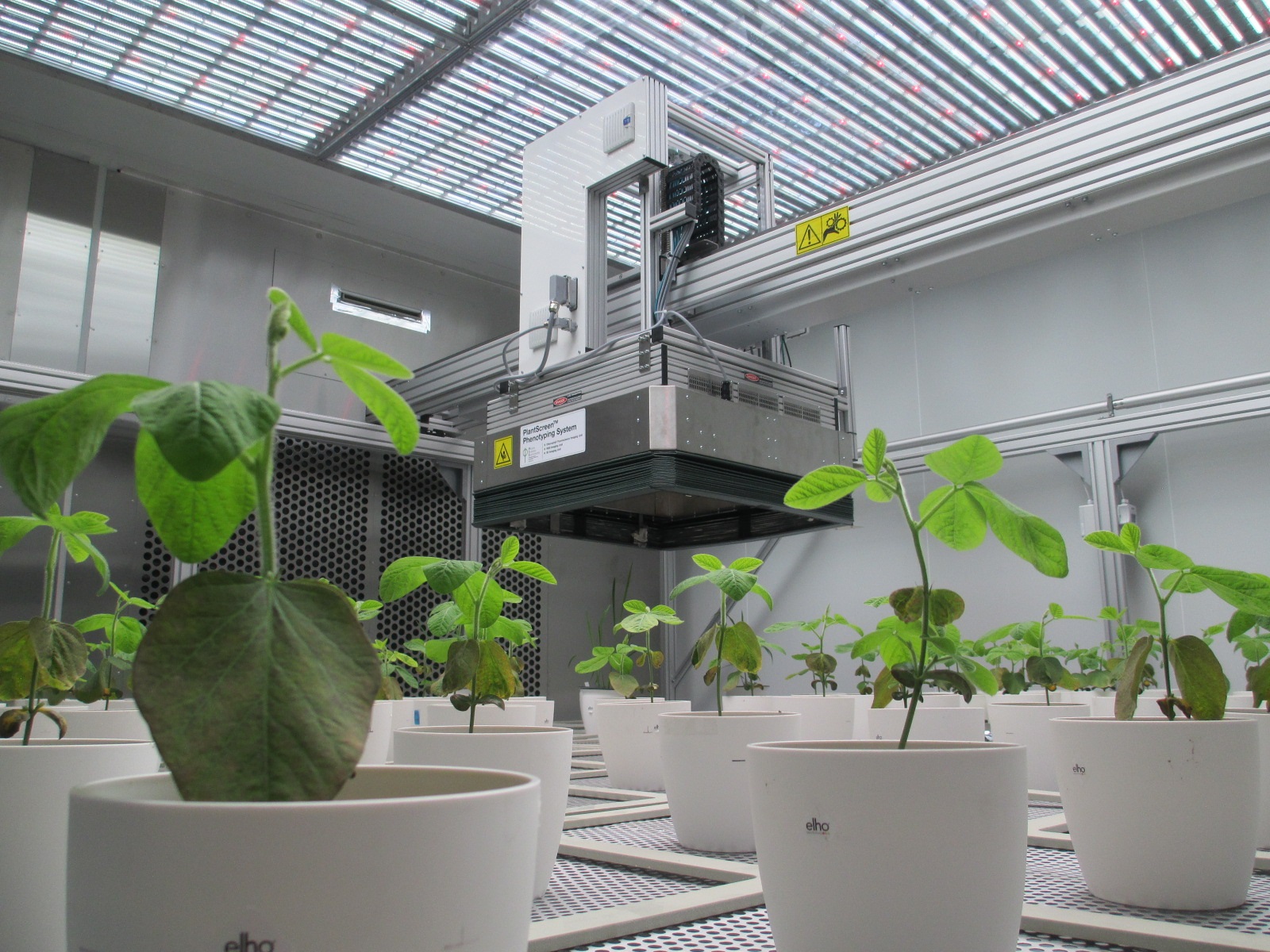
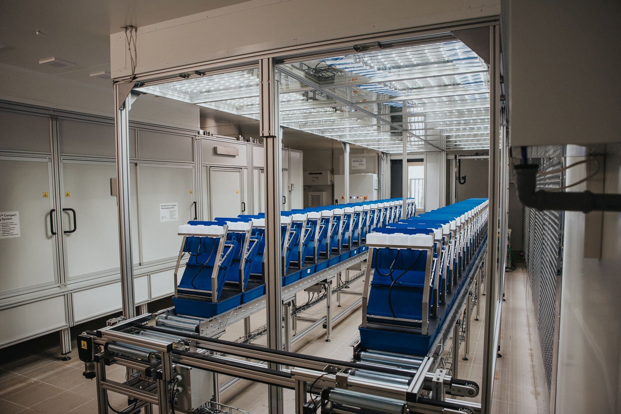
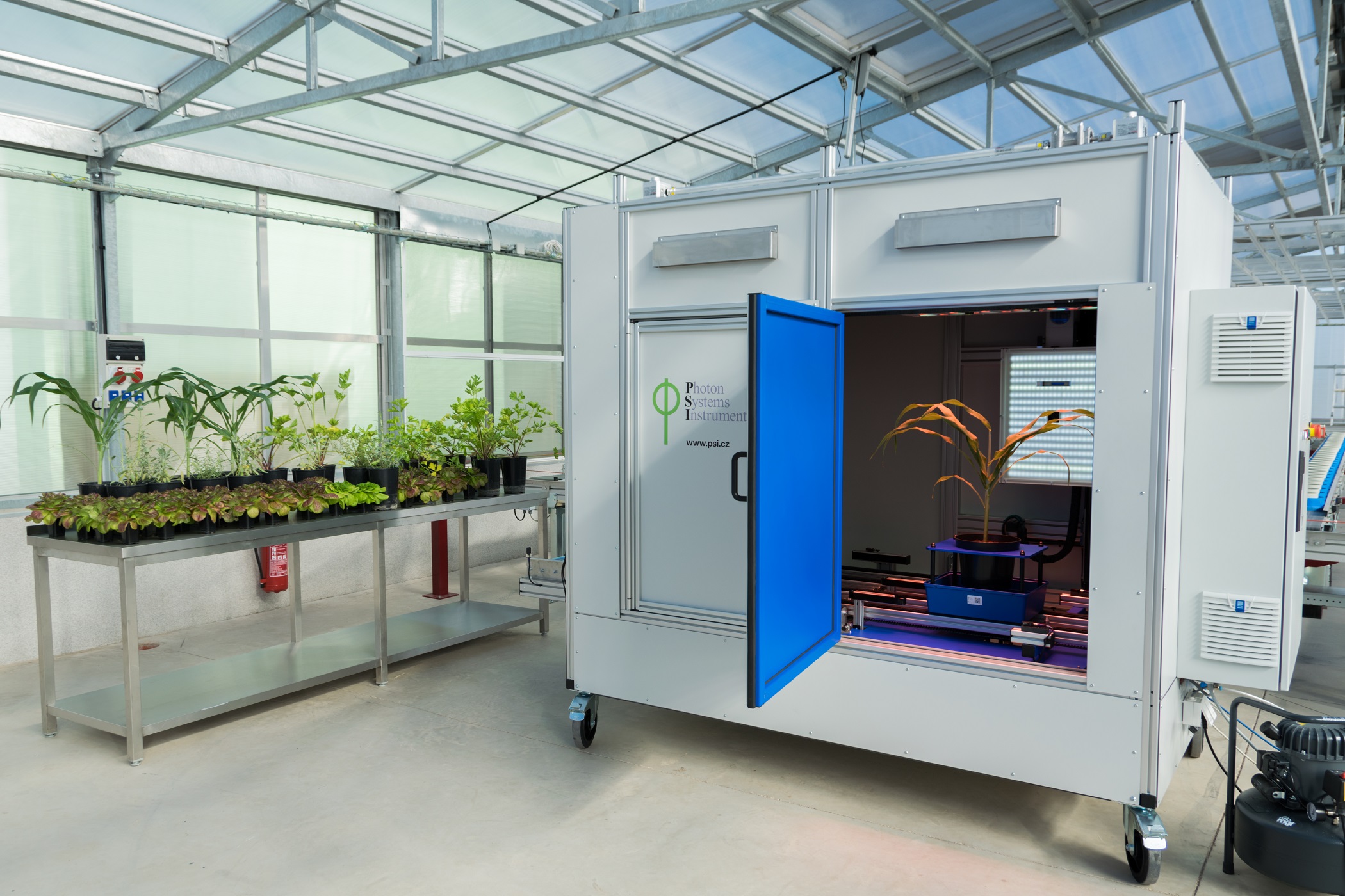
PlantScreen TM Imaging Sensors are complete integrated solutions for non-invasive analysis of plant specific patterns of absorption, emission and reflection. Digital trait asses...
广西科米瑞实验仪器设备有限公司提供 Photon Systems Instruments (PSI) PlantScreen Imaging sensors 产品资料、选型咨询、供货报价和售后服务支持,价格面议。
← 返回产品列表
说明
PlantScreen TM Imaging Sensors are complete integrated solutions for non-invasive analysis of plant specific patterns of absorption, emission and reflection. Digital trait assesment sensors are based on analysis of wide range of electromagnetic radiation wavelength bands: visible range of spectra is detected by RGB cameras for structural and color analysis, hyperspectral cameras in visible, near-infrared and short wavelength infra-red range of spectra are used for reflectance-based analysis of plants, thermal imaging cameras are optimized for leaf temperature and stomata conductance analysis, kinetic chlorophyll fluorescence imaging sensors for analysis of plant photosynthetic performance.
Range of high-end sensors
Sensor-specific light sources
Light-isolated imaging box
Sensor-specific raw data processing software
Comprehensive software package
RGB and Morphometric Imaging Wide range number of features linked to plant growth and development can be extracted from digital colour RGB imaging or 3D scanning technology, when connected to the automatic software analysis. RGB digital imaging applied in high resolution is used for in-depth analysis of plant morphology, architecture and colour index analysis.
Industrial high performance cameras with a Gbit Ethernet connection are mounted on robotic arm together with the white LED light source to ensure high speed data transfer and precise color separation. Cameras with high-sensitivity CCD-sensors, high-resolution and broad dynamic range are used.
Key features 2D or 3D scanning mode Top view up to 150 cm plant height Side view up to 150 cm plant height Side view in range 0-360° Line scanning mode for side view Species oriented analysis (mono and di-cotyledonous plants) Static and dynamic analysis Homogenous LED light source
Top view up to 150 cm plant height
Side view up to 150 cm plant height
Side view in range 0-360°
Line scanning mode for side view
Species oriented analysis (mono and di-cotyledonous plants)
Static and dynamic analysis
Homogenous LED light source
Download the information brochure (pdf)
Kinetic chlorophyll fluorescence Imaging Chlorophyll fluorescence is popular technique in plant physiology used for rapid non-invasive measurement of photosystem II (PSII) activity. PSII activity is very sensitive to range of biotic and abiotic factors and therefore chlorophyll fluorescence technique is used as rapid indicator of photosynthetic performance of plants in different developmental stages and/or in response to changing environment. Systems developed by PSI monitor fluorescence kinetics in pulse-amplitude modulated mode , which provides a wealth of information about a plant's photosynthetic capacity, physiological and metabolic condition, as well as its susceptibility to various stress conditions.
The advantage of chlorophyll fluorescence measurements over other methods for monitoring stresses is that changes in chlorophyll fluorescence kinetic parameters often occur before other effects of stress are apparent. The method is non-invasive, and the spread of inhibition can be observed and quantified with time. Heterogeneity in the location of inhibition is easily seen and quantified when using imaging systems to measure chlorophyll fluorescence. Chlorophyll fluorescence kinetics measured in the PlantScreen TM Phenotyping Systems provides a wealth of information about a plant's photosynthetic capacity, physiological and metabolic condition, as well as its susceptibility to various stress conditions.
Key features High sensitivity CCD camera Multi-colour LED light panel Pulse-modulated short duration flashes for accurate measurement of minimal fluorescence (Fo value) determination Two types of actinic lights for light-adapted and quenching analysis with maximum light intensity reaching 2000 µmol.m -2 .s -1 . Saturating light pulse for maximal fluorescence Fm value determination with maximal light intensity up to 6000 µmol.m -2 .s -1 Additional light: FAR (735 nm) for determination of Fo'. Royal blue (450 nm) as excitation light source for GFP detection. Camera includes 7 position filter wheel, ChlF and GFP filter and other filter sets according to user's needs. Custom-designed open FluorCam to image plant in trays and pots of various dimensions with maximum imaged area of 80 cm x 80 cm User defined programmable measuring protocols. Automatic data analysis and parameters computation
High sensitivity CCD camera
Multi-colour LED light panel
Pulse-modulated short duration flashes for accurate measurement of minimal fluorescence (Fo value) determination
Two types of actinic lights for light-adapted and quenching analysis with maximum light intensity reaching 2000 µmol.m -2 .s -1 .
相关产品
提交需求
请填写必填信息,我们将在工作日尽快回复。